Executive Summary
Hindgut ulcers in horses, also referred to as equine hindgut ulcers, colonic ulcers in horses, or cecal ulcers in horses, represent one of the most clinically important yet frequently overlooked forms of hindgut disease in horses. While awareness of gastric ulcers has improved, hindgut ulcers affecting the large intestine continue to be under-recognized, despite strong evidence that they are closely linked to chronic digestive discomfort, performance decline, and behavioral changes. Modern research in equine hindgut health shows that these lesions rarely exist in isolation. Instead, they arise from hindgut fermentation in horses becoming unstable, leading to hindgut acidosis in horses, microbial imbalance, altered metabolite output, and progressive compromise of the gut lining.
A central mechanism identified in deep research is disruption of short-chain fatty acid availability, particularly butyrate deficiency, which directly weakens colonocyte energy supply and mucosal resilience. When feeding practices, stress, dehydration, or NSAID gut damage in horses interfere with normal fermentation, the result can be hindgut inflammation in horses, intestinal permeability, and ulcer formation. Horses affected by this process often show free fecal water syndrome in horses, intermittent colic, girthy behavior linked to gut pain, or poor performance related to digestive dysfunction. This article explains what causes hindgut ulcers in horses, why they are often missed, and how early recognition combined with nutrition-driven prevention strategies can protect long-term equine digestive health.
Introduction
A horse that has always been reliable may suddenly become girthy, resistant under saddle, or unusually sensitive during grooming. Another may experience intermittent colic in horses that resolves quickly but recurs without warning. Others show a gradual decline in performance, unexplained weight loss, or persistent free fecal water syndrome (FFWS) in horses, even though the feed program appears unchanged. These scenarios are common across disciplines and are often attributed to training, temperament, or vague digestive upset.
In many cases, attention immediately shifts to the stomach, and gastric ulcers are suspected. Yet when treatment fails to fully resolve the problem, the underlying issue may lie deeper in the digestive tract. The purpose of this article is to explain hindgut ulcers vs gastric ulcers in horses, clarify why equine colon irritation and large intestine ulcers in horses are frequently overlooked, and demonstrate how recognizing hindgut ulcer symptoms early can prevent long-term digestive damage. By understanding how feeding, fermentation, and stress interact in the hindgut, readers can improve comfort, consistency, and performance while reducing the risk of chronic digestive disease.
What Are Hindgut Ulcers?
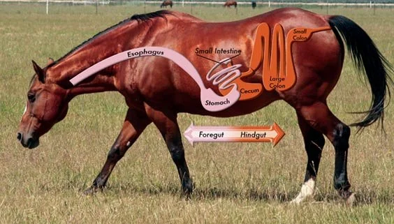
Hindgut ulcers are erosive or ulcerative lesions affecting the mucosa of the cecum and large colon. In clinical reality, equine colonic ulcers and cecal inflammation in horses rarely appear as isolated findings. They commonly coexist with hindgut dysbiosis in horses, inflammation of the equine large intestine, and functional signs of equine gastrointestinal dysfunction.
From a practical perspective, hindgut ulcers can be understood as a form of hindgut mucosal damage and intestinal barrier failure. The large intestine is designed to absorb nutrients while preventing harmful fermentation byproducts from entering circulation. When this barrier is repeatedly stressed, gut lining damage in horses develops, increasing vulnerability to ulceration and systemic inflammatory effects.
Hindgut Ulcers vs. Gastric Ulcers
Both conditions fall under the broader category of ulcers in horses, yet their underlying mechanisms differ. Gastric ulcers are driven primarily by stomach acid exposure and localized mucosal defense failure. In contrast, horse hindgut ulcers develop in a fermentation-dependent environment where starch overload in horses, microbial activity, hydration status, and stress physiology determine risk.
This distinction has important clinical implications. Acid-suppressive strategies effective for gastric ulcers do not address starch spillover and hindgut acidosis in horses, lactic acid buildup in the hindgut, or epithelial energy deficits in the colon. As a result, horses with hindgut ulcer syndrome may show partial improvement followed by relapse when the hindgut remains unaddressed.
The Hindgut as a Metabolic and Immune Organ
The hindgut is not simply a waste-processing organ. It is a complex metabolic system responsible for fiber digestion in horses through microbial fermentation. This process generates short-chain fatty acids, including acetate, propionate, and butyrate.
Among these metabolites, butyrate is particularly critical. SCFA deficiency in horses, especially butyrate deficiency, compromises colonocyte energy supply, tight junction integrity, and inflammatory control. Deep research highlights that when fermentation becomes unstable and metabolite output shifts, intestinal barrier dysfunction in horses develops, linking hindgut disease to immune activation, systemic inflammation, and even the gut-immune axis in horses.
Why the Hindgut Is Especially Vulnerable
Unlike the stomach, the hindgut lacks robust buffering mechanisms. Its stability depends on continuous fiber intake, consistent microbial populations, and adequate hydration. Feeding gaps in horses, irregular hay feeding schedules, and dehydration can rapidly destabilize fermentation.
Repeated exposure to these stressors leads to fermentation imbalance in horses, low hindgut pH, and progressive mucosal injury. Over time, this cumulative damage increases the likelihood of hindgut ulcers and poor performance in horses.
Primary Causes of Hindgut Ulcers
One of the most common drivers of hindgut ulcers is dietary starch spillover. When a high grain diet in horses exceeds small-intestinal digestive capacity, undigested starch reaches the hindgut. Rapid fermentation follows, producing lactic acid and promoting equine hindgut acidosis.
This acidic environment suppresses fiber-fermenting microbes, reduces beneficial SCFA production, and directly irritates the colonic lining. Over time, repeated episodes of acidification and recovery contribute to hindgut inflammation, colonic mucosal injury in horses, and ulcer formation.
Forage disruption is another major factor. Forage-first feeding in horses—supported by consistent access to long-stem forage—helps stabilize fermentation and protect the hindgut. Long periods without forage, inadequate fiber intake, or poorly managed slow feeder hay nets for horses increase ulcer risk.
Symptoms Often Mistaken for Other Problems
The symptoms of hindgut ulcers in horses are often subtle and easily misattributed. Common signs include chronic mild colic in horses, fluctuating manure consistency, and free fecal water syndrome and colic in horses occurring together.
Other indicators include unexplained weight loss, dull coat, reduced appetite, and poor performance in horses related to digestive pain. Behavioral signs are especially misleading. Behavior changes from gut pain in horses may present as girthiness, resistance to bending, irritability during grooming, or reluctance to work, frequently mistaken for training or orthopedic issues.
A key practical insight is persistence. When digestive or behavioral signs continue despite reasonable gastric management, clinicians should consider hindgut dysfunction in horses more seriously.
Diagnostic Challenges and Practical Decision-Making
Direct visualization of the hindgut is rarely feasible, making diagnosis challenging. As a result, many professionals rely on a systems-based approach to how to detect hindgut ulcers in horses. This includes evaluating feeding practices, starch exposure, forage availability, hydration, workload, travel stress, and medication history.
A response-to-intervention strategy is commonly used. When targeted adjustments—such as reducing starch load, improving forage access, and stabilizing hydration—lead to improvements, the clinical picture becomes consistent with hindgut ulcer treatment without scoping. This approach does not replace veterinary diagnostics but provides a practical pathway for early intervention.
Feeding and Management Solutions
Effective management focuses on stabilizing fermentation and protecting the gut lining. The best feeding practices for hindgut ulcers in horses emphasize consistency, fiber availability, and controlled starch exposure.
The best diet for horses with hindgut ulcers is built around forage-first principles. Continuous access to quality forage supports stable fermentation and SCFA production. Concentrate meals should be evaluated by starch load per meal rather than total daily intake. The question does grain cause hindgut ulcers in horses is best answered by recognizing that excessive starch delivery—not grain itself—is the primary risk factor.
Where additional calories are required, fiber-based energy sources such as beet pulp for horses’ gut health and dietary fats are often preferred to reduce fermentation volatility. Hydration management is equally important, as adequate water intake supports normal transit and reduces mucosal stress.
Stress governance and medication review also play critical roles. Reducing unnecessary bute side effects on the horse gut, planning feeding routines during travel, and allowing adequate recovery after intense work all contribute to improved hindgut stability.
Scientifically Backed Prevention Strategies
Prevention focuses on minimizing repeated micro-insults to the hindgut. How to prevent hindgut ulcers in horses centers on consistent forage access, controlled starch intake, gradual dietary transitions, and reliable hydration.
From a metabolome perspective, these practices preserve SCFA production, protect against metabolome disruption in horses, and maintain epithelial energy supply. Routine monitoring of manure quality, behavior, and performance helps identify early signs of hidden gut issues in horses before ulcers develop.
Conclusion
Hindgut ulcers in horses are a hidden but clinically significant contributor to chronic digestive issues, behavioral resistance, and performance decline. They develop gradually as gut microbiome imbalance in horses, fermentation instability, and barrier compromise accumulate over time. Because signs are subtle and often mistaken for other problems, early recognition is essential. By understanding the hindgut as a metabolic system rather than an isolated organ, professionals can address root causes and support long-term digestive resilience.
Frequently Asked Questions (FAQs)
Q1:What causes hindgut ulcers in horses?
A: Hindgut ulcers develop when repeated starch overload, forage gaps, stress, dehydration, or NSAID exposure disrupt fermentation, leading to hindgut acidosis, microbial imbalance, and mucosal injury.
Q2: Are hindgut ulcers painful for horses?
A: Yes. Although pain may be subtle, hindgut ulcers can cause chronic digestive discomfort, behavioral changes, and poor performance.
Q3: Can hindgut ulcers cause laminitis?
A: Hindgut ulcers can contribute to systemic inflammation and endotoxemia in the hindgut, increasing laminitis risk in susceptible horses
Call to Action
If your horse shows recurring colic-like signs, persistent free fecal water, hindgut-related behavior issues, or unexplained performance decline, take a closer look at hindgut health. Review forage access, starch exposure, hydration, stress load, and medication history with your veterinarian. Early, informed action can prevent progression and improve comfort, performance, and long-term wellbeing.
References
- Andrews, F. M., Buchanan, B. R., Elliott, S. B., Clariday, N. A., & Edwards, L. H. (2005). Results of a large-scale necroscopic study of equine colonic ulcers. Journal of Equine Veterinary Science, 25(3), 113–117.
- Flood, J., et al. (2023). Right dorsal colitis in horses: A multicenter retrospective study of 35 cases. Veterinary Medicine and Science, 9(1), e1234.
- Hamer, H. M., Jonkers, D., Venema, K., Vanhoutvin, S., Troost, F. J., & Brummer, R. J. (2008). The role of butyrate on colonic function. Alimentary Pharmacology & Therapeutics, 27(2), 104–119.
- Julliand, V., & Grimm, P. (2017). The microbiome of the horse hindgut: History and current knowledge. Animal Frontiers, 7(3), 55–59.
- Laustsen, L., et al. (2021). Free fecal water syndrome in horses: Associations with microbiota and management. Animals, 11(5), 1321.
- Merritt, A. M., & Julliand, V. (2013). Gastrointestinal disorders of horses. In Equine Internal Medicine (3rd ed.). Saunders.
- Murray, M. J. (2013). Equine gastric ulcer syndrome. Journal of Veterinary Internal Medicine, 27(2), 219–232.
- National Research Council. (2007). Nutrient requirements of horses (6th rev. ed.). National Academies Press.
- Sykes, B. W., Hewetson, M., Hepburn, R. J., Luthersson, N., & Tamzali, Y. (2015). European College of Equine Internal Medicine consensus statement—Equine gastric ulcer syndrome. Journal of Veterinary Internal Medicine, 29(5), 1288–1299.
- Whitfield-Cargile, C. M., et al. (2021). Intestinal injury, permeability, and repair in horses: Emerging concepts. Veterinary Clinics of North America: Equine Practice, 37(2), 311–329.